Here are some of the observation results using NanoSuit technology.
GalleryII (Example using Type II)
●Charge reduction effect of NanoSuit solution
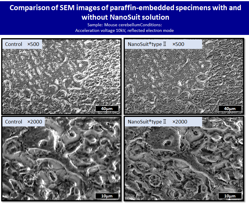
Left) Without NanoSuit solution, glare and distortion of the image are observed due to charge-up. Also, it gets worse as the observation magnification is increased.
Right) In the image using NanoSuit® solution type II, the charge-up is reduced and a fine structure is observed.
●Bacterial infection
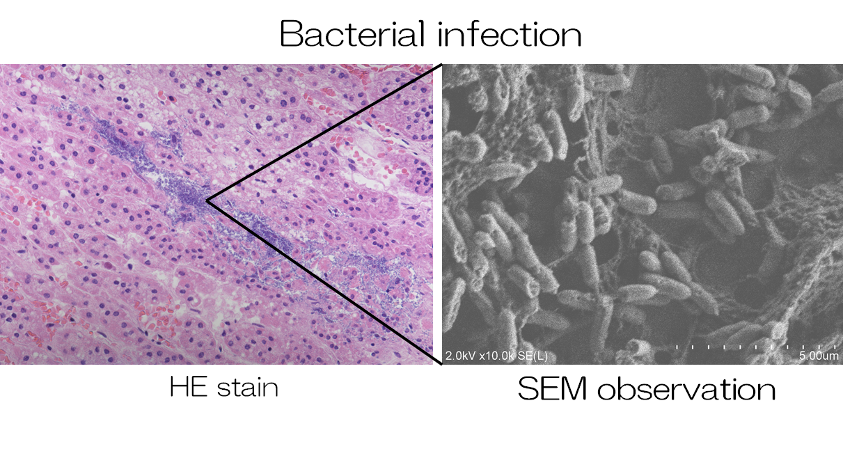
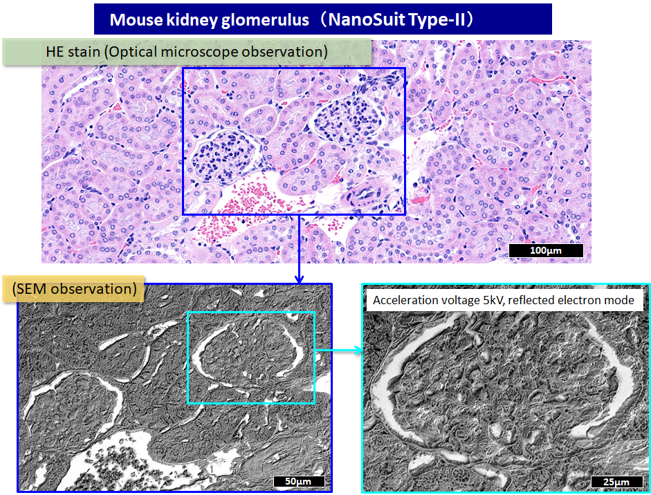
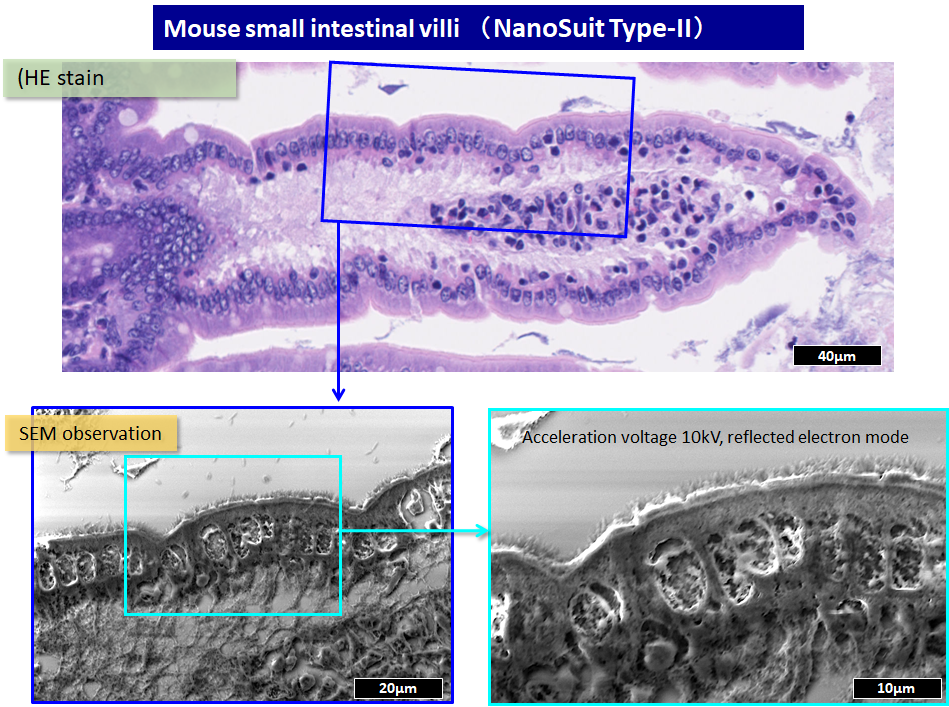
By coating the area where the stained image is obtained with an optical microscope with NanoSuit Type II and then observing it with an electron microscope (SEM), you can magnify and obtain a three-dimensional image. In this case, the presence of bacteria can be clearly observed.
●Bronchiole pili of mouse lung
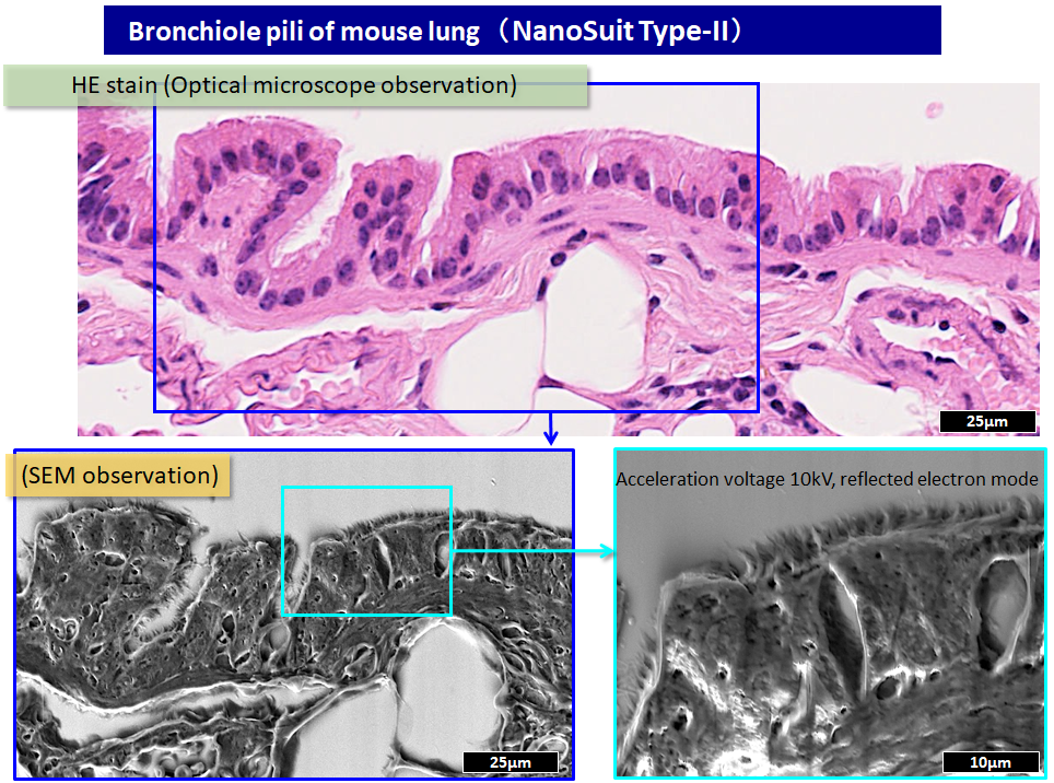
After observing the HE-stained sample with an optical microscope, it was observed with a scanning electron microscope using NanoSuit solution Type II. Fimbria was observed as it was magnified with an electron microscope.
●Mouse kidney glomerulus
●Mouse small intestine
●Helicobacter pylori
